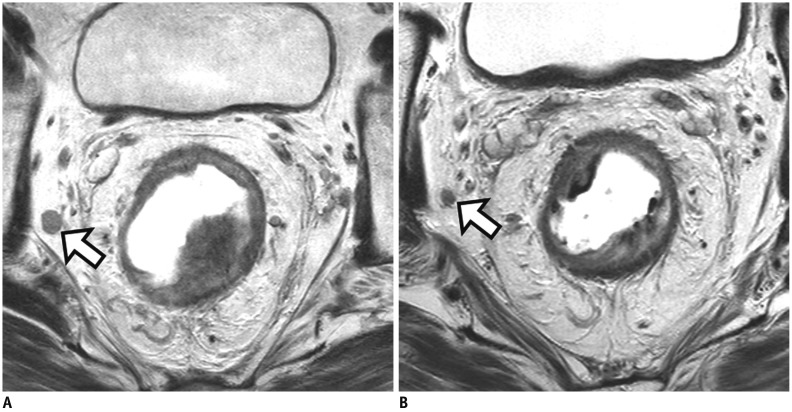
Fig. 9. Tumor response of lymph node after CRT.
A. On baseline MRI, suspicious metastatic node with short axis diameter of 9 mm is seen at right lateral pelvic side wall (arrow). B. Post-CRT MRI reveals interval decrease of node (arrow) and size is less than 5 mm. Lymph node dissection at right lateral pelvic side wall was performed. Viable tumor cells were not found in dissected lymph nodes (not shown). However, pathologic nodal status after CRT still remains uncertain because both normal and metastatic lymph nodes show interval decrease in size after CRT.

