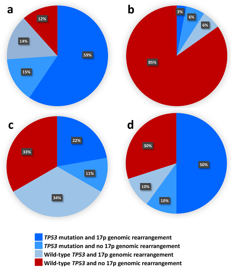
Figure 2.

Distribution of the combinations of TP53 mutations and 17p genomic rearrangements (determined using SNP arrays) in subsets of patients with acute myeloid leukemia (AML) and a complex karyotype (CK): (a) typical CK (n=69), (b) atypical CK (n=33), (c) CK with rare recurrent balanced chromosome abnormalities (n=9), (d) CK with unique balanced chromosome abnormalities (n=10). Dark blue color denotes patients with both TP53 mutation and 17p genomic rearrangement; lighter blue, patients with TP53 mutation and no 17p genomic rearrangement; light blue, patients with wild-type TP53 and 17p genomic rearrangement present, and red color indicates patients with wild-type TP53 and no 17p genomic rearrangement. All patients in the first three subsets combined, indicated by the various shades of blue, are considered to harbor an alteration of TP53 (i.e., TP53 mutation, deletion of 17p resulting in loss of TP53 locus and/or copy-neutral loss of heterozygosity encompassing TP53 locus). Typical CK-AML (a) clearly differs from atypical CK-AML (b) with regard to the frequency of TP53 alterations (88% vs 15%; P<0.001).
