
Fig. 3.
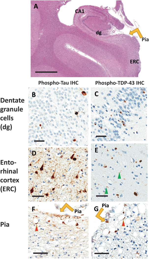
Comorbid Tau and TDP-43 pathologies are relatively common pathologic phenomena. Here are shown photomicrographs depicting stained sections from the brain of a 102-year-old woman who died with a history of dementia. Autopsy showed minimal Alzheimer’s disease-type changes (no neuritic Aβ plaques and Braak NFT stage II). Portions of the brain were stained using hematoxylin and eosin (H&E; a), and near-adjacent sections were stained for phospho-Tau immunohistochemistry (IHC; b, d, f), and phospho-TDP-43 IHC (c, e, g). The hippocampus is shown in the coronal plane (a) with anatomic regions labeled. Panels b and c show dentate granule (dg) cells, d and e show entorhinal cortex (ERC), and f and g demonstrate IHC staining near the pia lining (orange arrows). Sections are counterstained using hematoxylin (blue nuclei) and IHC reaction product is brown. Note that both Tau and TDP-43 proteinopathy are seen in dentate granule cells, entorhinal cortex, and within twiglike processes around corpora amylacea (red arrowheads) near the pia layer at the surface of the medial temporal lobes. Scale bars = 4 mm (a), 70 μm (b–e), and 100 μm (f, g)