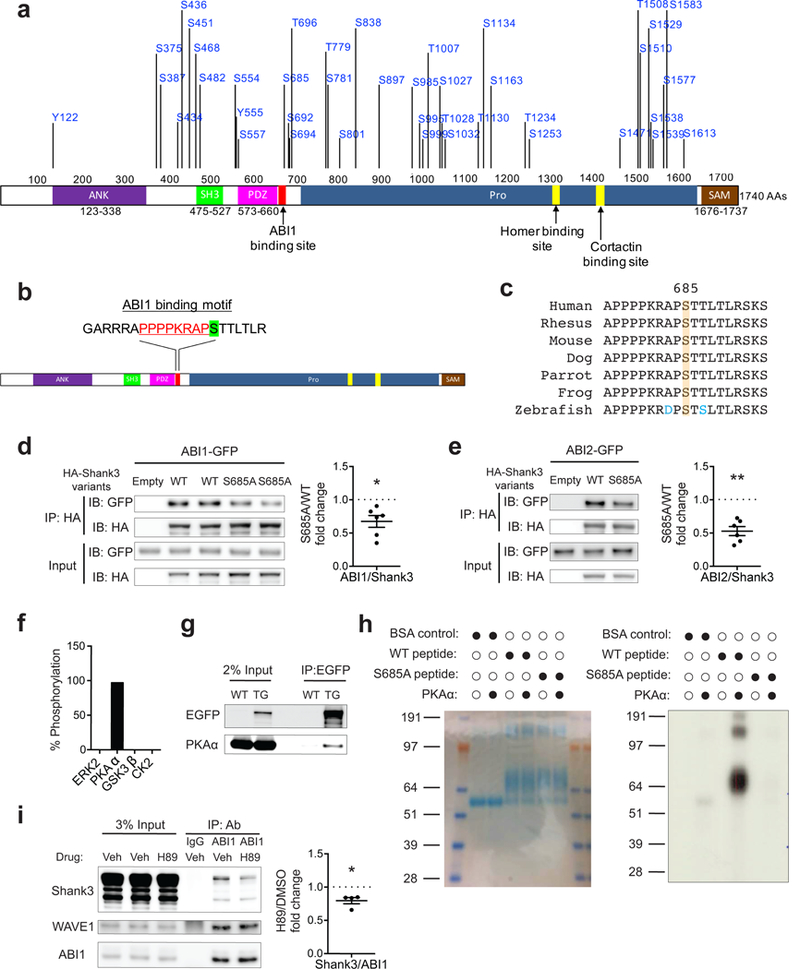
Figure 1.

S685 phosphorylation modulates Shank3-ABI interaction. (a) Diagram of Shank3 protein (NP_067398) with 41 in vivo phosphorylated residues identified by mass spectrometry. (b) Diagram of Shank3 protein with S685 (green highlighted) adjacent to a previously identified ABI1 binding site (red text) on Shank3. (c) Protein sequence alignment of Shank3 surrounding S685 among vertebrates. (d,e) Immunoblots of ABI1-GFP (d) or ABI2-GFP (e) and HA-Shank3 following immunoprecipitation of HA-Shank3 variants in the co-immunoprecipitation assay in 293T cells (n=6, n=6 experiments). (f) Percentage of peptides with S685 phosphorylation in Shank3 mass-spectrometry-based in vitro kinase assay by ERK2, PKAα, GSK3β, and CK2. (g) Immunoblots of PKA α-catalytic subunit following immunoprecipitation of EGFP-tagged Shank3 from EGFP-Shank3 transgenic mice striatal lysate. TG, EGFP-Shank3 transgenic mice. (h) In vitro kinase assay with radiolabeled ATP using recombinant PKAα and Shank3 peptides including S685. Left, coomassie blue staining of BSA-conjugated peptides (aa 668–691). Right, autoradiography blot of kinase reaction product. (i) Immunoprecipitation of ABI1 in primary cortical neurons treated with vehicle (DMSO) or H89 (10 μM) followed by immunoblotting for Shank3, WAVE1, and ABI1 (n=4 experiments). WAVE1-ABI1 co-immunoprecipitation was used as positive control. Veh, vehicle (DMSO). All data are presented as mean ± s.e.m. *p<0.05, **p<0.01; paired two-tailed Student’s t-test.
