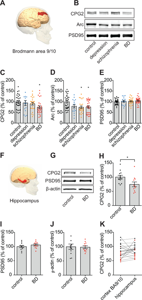
Figure 1: CPG2 protein levels are reduced in postmortem brain tissue from BD patients.
A) Synaptic protein fractions were extracted from a total of 102 postmortem human brain tissue samples from Brodmann Area 9/10 (image adapted from commons.wikimedia.org). For SDS-PAGE, 20µg of total protein was loaded. B) Representative Western blots comparing CPG2, Arc, and PSD95 protein expression in controls, depression, schizophrenia, and BD patients. Arc is another glutamatergic synaptic protein that like CPG2 is the product of an activity-regulated gene, and the synaptic protein PSD95 serves as a positive marker of glutamatergic synapse presence. C-E) Quantification of CPG2, Arc, and PSD95 protein levels, respectively, shows that CPG2 levels are significantly lower in the BD patient population as compared to control subjects, whereas Arc is decreased in both schizophrenia and BD groups. Comparable PSD95 levels in all groups indicates that reduced CPG2 or Arc expression does not reflect synaptic loss. (*p<0.05, One-way ANOVAs, Tukey’s post hoc tests). F) Synaptic protein fractions were extracted from a total of 22 postmortem human brain tissue samples from hippocampus (image adapted from commons.wikimedia.org). G) Representative Western blots showing CPG2 protein expression in controls and BD patients. The synaptic marker protein PSD95 and β-actin serve as controls. H) Quantification of CPG2 protein expression. I) Quantification of PSD95 protein expression. J) Quantification of β-actin protein expression. K) Direct comparison of CPG2 protein expression in cortical and hippocampal tissue samples from individual subjects (black dots represent controls and red dots represent BD patients). As in neocortex, CPG2 protein expression is significantly decreased in hippocampal tissue from BD patients. (*p<0.05, Student’s t-tests).

