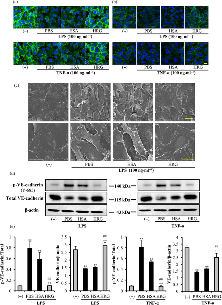
Figure 2.

Histidine‐rich glycoprotein (HRG) prevents the loss of intercellular junction. Immunostaining results of VE‐cadherin/β‐catenin in endothelial cells stimulated with 100 ng·ml−1 LPS or TNF‐α for 6 hr after treatment with HRG or human serum albumin (HSA). (a, b) Cells were incubated with anti‐VE‐cadherin mAb or anti‐β‐catenin mAb for 2 hr and then stained with Alexa Fluor 488 (green) goat‐anti‐rabbit IgG. The cells were also stained with DAPI (blue) to visualize the nuclei. The pictures are representative of three independent experiments (n = 5 per group). Scale bar = 20 μm. (c) Scanning electron microscopy pictures of vascular endothelial cells. Scale bar = 50 μm (upper panel) or 20 μm (lower panel). (d, e) Quantification of results of the p‐VE‐cadherin and VE‐cadherin by Western blotting. One‐way ANOVA followed by the post hoc Fisher test, **P < .05 versus control, ## P < .05, and ++ P < .05 versus PBS and HSA respectively
