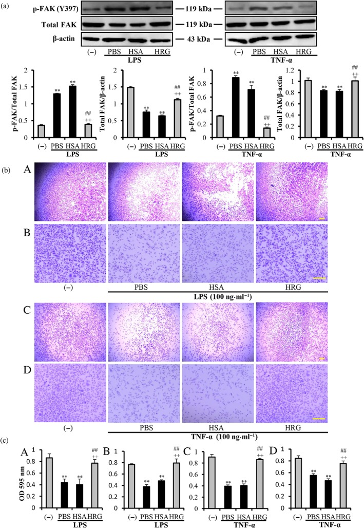
Figure 3.

Effects of histidine‐rich glycoprotein (HRG) on the degradation of focal adhesion kinase (FAK) and cell detachment. (a) Western blot results and quantification of the phosphorylated FAK (p‐FAK) and degraded FAK induced by LPS/TNF‐α with ImageJ. (b) The cell suspensions were plated into 96‐well plain plates (A, C) or laminin‐coated plates (B, D). The cell detachment was evaluated by staining the remaining cells with 1% crystal violet. (c) Quantitative analysis of crystal violet extracted from the remaining cells. Panels A–D correspond to A–D in (b). Data are means ± SEM in the graph (n = 5 per group). One‐way ANOVA followed by the post hoc Fisher test, **P < .05 versus control, ## P < .05, and ++ P < .05 versus PBS and human serum albumin (HSA) respectively. Scale bar = 10 μm
