
Figure 5.
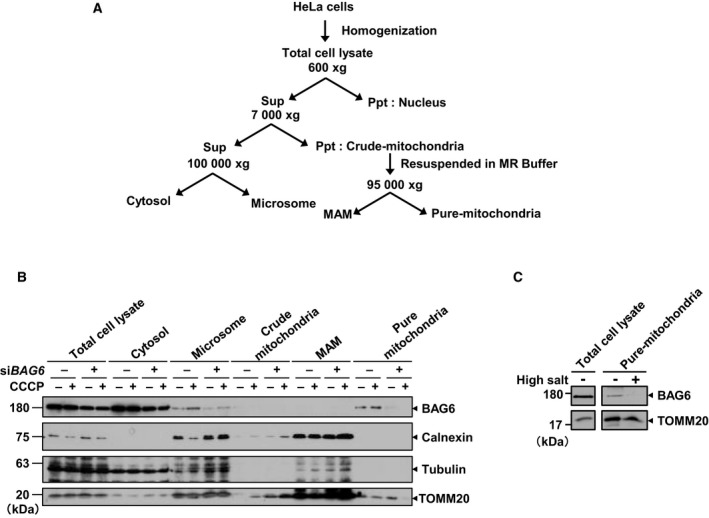
BAG6 was localized in the mitochondrial fraction. (A) Schematic diagram of the fractionation protocol. (B) Fractions were analyzed by western blotting. Tubulin, calnexin, and TOMM20 were used as markers for the cytoplasm, microsome, and mitochondria, respectively. The grouping of blots cropped from different parts of the same membrane. (C) Pure mitochondrial fractions isolated from HeLa cells were incubated with high‐salt buffer 1 m KCl, 600 mm mannitol, 20 mm HEPES‐KOH, pH 7.5, 2 mm MgCl2, and 1 mm PMSF) for 15 min on ice and then centrifuged for 5 min at 10 000 g. The resultant high‐salt‐washed pure mitochondria were recovered as a pellet. The samples were separated by sodium dodecyl sulfate‐polyacrylamide gel electrophoresis and immunoblotted for BAG6 and TOMM20, which was used as a control for mitochondria‐embedded protein.