Figure 9.
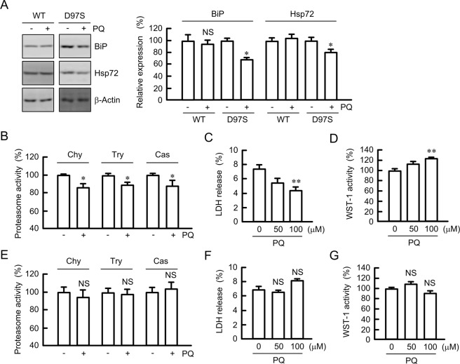
Decrease in chaperone protein, proteasome activity, and cell injury by primaquine. (A) The FLAG-tagged WT CLDN16- or D97S mutant-expressing cells were treated with primaquine (PQ, 100 μM) for 6 h. After collecting cell lysates, the aliquots were blotted with anti-BiP, anti-Hsp72, and anti-β-actin antibodies. The band densities of proteins are represented relative to the values without PQ. The full-length blot images are shown in Supplementary Fig. S11. (B,E) The cytosolic fraction was isolated from D97S mutant-expressing cells (B) or WT CLDN16-expressing (E) using a passive buffer. Chymotrypsin-like (Chy), trypsin-like (Try), and caspase-like (Cas) proteasome activities were measured using each selective substrates. (C,D,F and G) MDCK cells expressing the FLAG-tagged D97S mutant (C,D) or WT CLDN16 (F,G) were incubated with 100 μM PQ for 24 h. Cell injury was estimated by LDH release into the media and WST-1 activity. n = 4 in four independent experiments. **P < 0.01, *P < 0.05, and NS P > 0.05 compared with 0 h or without PQ.

