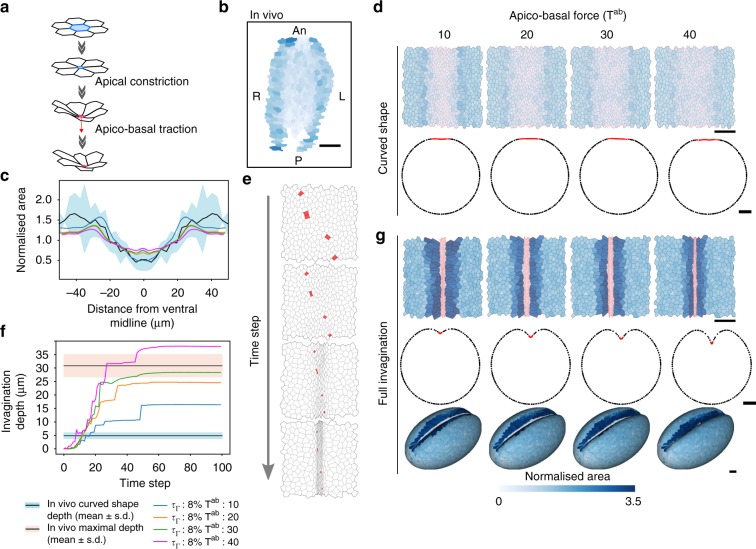
Fig. 8.
Apico-basal traction is required for mesoderm invagination in a 3D biophysical model. a Scheme of the apical constriction and the apico-basal traction processes. First, the cell constricts its apex (blue) until its surface area falls under a critical threshold. Then, the cell exerts an apico-basal force (red arrow) perpendicular to the apical surface. b Same as Fig. 4c. Central region of the curved-shaped mesoderm in vivo. Apical area is color-coded (see color bar in g). c Comparison of cell area in the curved-shaped mesoderm in vivo and in the model. Graphs show the normalized cell area in vivo (mean: black line, s.d.: blue band, n = 3) and in simulations for different values of apico-basal force Tab (color-coded as indicated in f) with respect to the distance from the ventral midline. d Top row: Images show the central region of the curved-shaped mesoderm in simulated embryos for different values of apico-basal force Tab and an apical contractility increase rate τΓ = 8%. Apical area is color-coded and cell edges are red in the mesoderm and black elsewhere (see color bar). Bottom row: Tissue deformation of the modeling results of curved-shaped embryos. For each simulation, curved shape was defined when the invagination depth corresponds to the in vivo depth of curved-shaped embryos. Scale bar: 25 µm. e Time-lapse images of the ventral part during mesoderm invagination showing the apical extrusion of a few cells (marked in red). f Invagination depth as a function of time for different values of apico-basal force Tab (color-coded as indicated below) and an apical contractility increase rate τΓ = 8%. The black line with a blue (resp. pink) border denotes the average depth measured in vivo when the embryo adopts a curved shape (resp. at the end of invagination). g Same as (d) at full invagination stage for upper and middle rows. Bottom: Tissue deformation (3D view) of the simulation results at full invagination stage. Scale bar: 25 µm See also Supplementary Fig. 4 and Supplementary Movie 9

