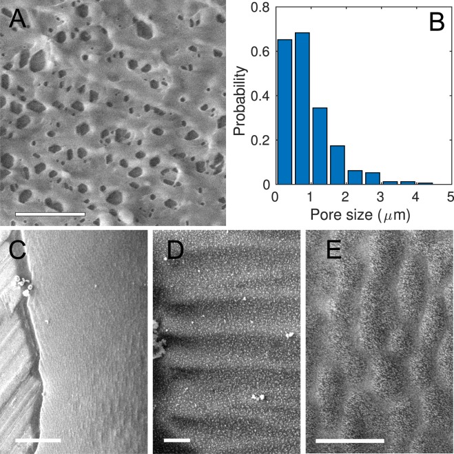
Figure 7.
Cryo SEM images of mucus. (A) Image of a drop showing a pore mesh. Scale bar 10 μm. (B) Histogram of pore sizes in mucus, as extracted from panel (A). (C) Mucus thread deposited vertically. Scale bar 100 μm. There is a vertical alignment of the mucus micro-structure, parallel to the orientation of the thread. The borders, however, show wrinkles almost perpendicular to the main direction. (D) Wrinkles at the edge of the thread, oriented perpendicular to the walls. Scale bar 10 μm. (E) Blowup of (C) showing the main vertical alignment close to the center of the thread. Scale bar 10 μm. Some ice crystals are present in panels (C) and (D).

