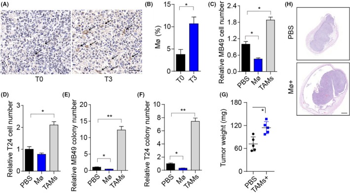
Figure 1.

Tumor‐associated macrophages (TAMs) promote bladder cancer cell growth and tumor progression. A, Immunohistochemistry of CD68 in bladder tumor tissues from patients in stage T0 and T3. Scale bar, 50 μm. The arrows indicate the macrophages in tumor sites. B, Percentage of macrophages in the immune cell subpopulation of patients (stage T0 and T3) bladder tumor tissues. C, Relative cell number of MB49 cells cocultured with mouse peritoneal macrophages, TAMs, or PBS for 72 h. D, Relative numbers of T24 cells cocultured with macrophages from patients’ paracarcinoma tissues, TAMs, or PBS for 72 h. E, Relative colony numbers of MB49 cells pretreated with mouse peritoneal macrophages, TAMs, or PBS for 72 h. F, Relative colony numbers of T24 cells pretreated with macrophages from patients’ paracarcinoma tissues, TAMs, or PBS for 72 h. G, Bladder tumor weights of mice treated with PBS or TAMs. Of note, 106 MB49 cells were intravesically instilled into C57 mice bladders. On days 4 and 8, mice were instilled with 2 × 10⁵ TAMs by bladder irrigation. H, Histological H&E staining of the bladders of C57 mice in PBS or TAM. Scale bar, 500 μm. Error bars, mean ± SEM; *P < .05; **P < .01
