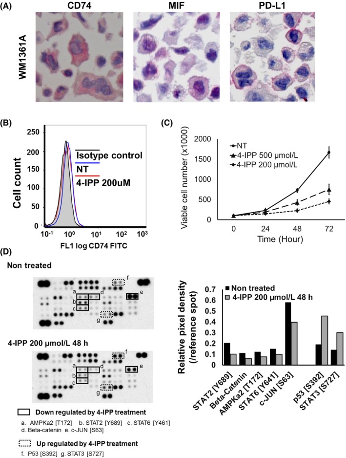
Figure 4.

Macrophage migration inhibitory factor (MIF)‐CD74 interaction regulates activity of several signaling molecules in the WM1361A melanoma cell line. A, Immunocytochemical analysis. WM1361A cells were confirmed to express programmed cell death ligand 1 (PD‐L1) (left panel), MIF (middle panel), and CD74 (right panel) in untreated cultures. B, Flow cytometry analysis. WM1361A cells showed cell surface expression of CD74. Mean fluorescence intensity of each condition was as follows. Isotype control, 0.47; nontreated (NT), 0.71; 4‐iodo‐6‐phenylpyrimidine (4‐IPP) 200 μmol/L, 0.65. C, Cell viability analysis. 4‐IPP treatment suppressed the proliferation of WM1361A cells in a dose‐dependent manner. D, Protein array. 4‐IPP treatment for 48 h decreased expression of phosphorylated (phospho‐)AMPKa2 (T172) (a), phospho‐STAT2 (Y689) (b), phospho‐STAT6 (Y461) (c), β‐catenin (d), and phospho‐c‐JUN (S63) (e), and increased expression of phospho‐P53 (S392) (f) and phospho‐STAT3 (S727) (g). Right panel, relative dot density of protein array analysis
