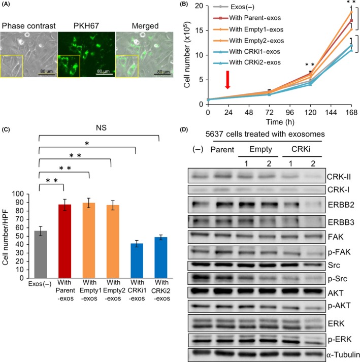
Figure 4.

Exosomes derived from high‐grade bladder cancer cells facilitate proliferation and invasion of low‐grade bladder cancer cells through an activation of ErbB2 signaling. A, 5637 cells (low‐grade) were treated with 20 μg PKH67‐labeled exosomes (green) derived from UM‐UC3 cells (high‐grade) for 48 h. Photomicrographs of the cells were obtained under bright‐field illumination (left) and using a fluorescence microscope (middle). B, Proliferation assay. The 5637 cells treated with exosomes derived from UM‐UC‐3 cells (parent, empty, and CRKi) (red arrow) were counted under a microscope at the indicated time points and expressed as the mean ± SD of 3 independent experiments. Cells treated with PBS were used as a control (Exos (−)). **P < .01 vs exos (−). C, Matrigel invasion assay. The 5637 cells were treated with exosomes derived from UM‐UC‐3 cells with or without CRK for 24 h, and the invading cells under the filter were counted and depicted as the mean ± SD. *P < .05 and **P < .01 vs Exos (−). D, In 5637 cells treated with UM‐UC‐3‐derived exosomes, expression and phosphorylation of the indicated proteins were examined by immunoblotting. FAK, focal adhesion kinase
