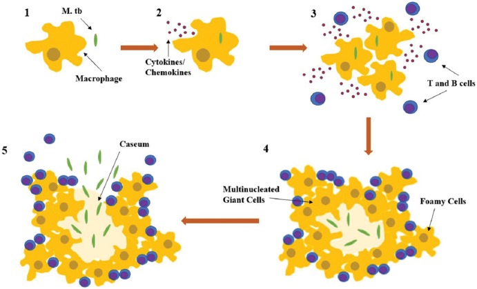
Fig. 2. Formation of the TB granuloma in primary lung infection and subsequent spread.
Following the inhalation of contaminated aerosols, Mycobacterium tuberculosis is recognized by macrophages in the lung alveoli by surface receptors (depicted in 1). Subsequently, the bacteria are taken up by macrophages which, along with epithelial cells and neutrophils, trigger innate immune signaling pathways that allow for the production of chemokines and cytokines (depicted in 2). The release of chemokines and cytokines recruits more macrophages, lymphocytes, and dendritic cells to the infection site, where they form granulomas composed of infected macrophages in the middle, surrounded by lymphocytes (CD4+, CD8+, gamma/delta T cells). The conglomerated macrophages can also fuse to form multinucleated giant cells or differentiate into lipid rich foamy cells (depicted in 3 and 4). Within the granuloma, bacteria can survive for years, in a latent disease state. However, once triggered by external factors, such as additional immunocompromising states, the bacteria can reactivate, killing the core infected macrophages and, thereby, producing a necrotic zone at the center of the granuloma known as a caseum (depicted in 4). The granulomatous structure weakens with the caseum and eventually breaks down, releasing bacteria through the body by blood, lymph, and infectious aerosolized droplets. Abbreviation: TB, tuberculosis.

