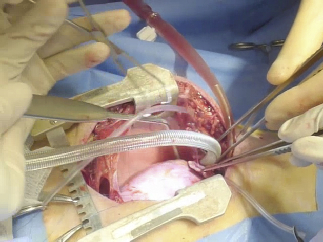
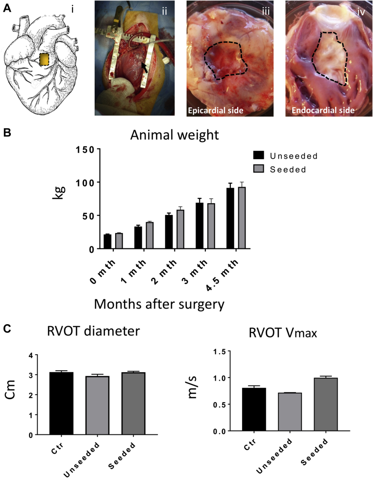
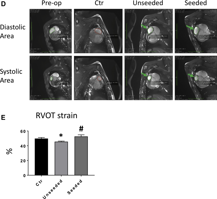
Figure 3.
in Vivo Right Ventricular Outflow Tract Reconstruction and Operated Animals Follow-Up
(A) Cartoon (i) and macroscopic image (ii) showing the site of the implant on the right ventricular outflow tract (RVOT). Gross analysis of the explants after 4.5 months in vivo shows the epicardial (iii) and endocardial (iv) sides of the graft (dotted line). (B) The operated pigs increased their body weight at a normal rate. (C) Doppler echocardiographic measurements of the right ventricle immediately before termination demonstrate comparable RVOT diameter and maximum velocity (Vmax) in the operated animals and unoperated control (Ctr) pigs. (D) Representative cardiac magnetic resonance images of right ventricular diastolic and systolic area (encircled in green) before surgery and 4.5 months thereafter. The patches could be visualized as a small bump protruding from the right ventricle (arrows). (E) RVOT myocardial strain measured at termination was greater in the animals implanted with seeded grafts. See Supplemental Video 1.


