
Figure EV3. Demonstration of neuronal degeneration in the cerebral cortex of mice subjected to permanent ischemia by photothrombosis.
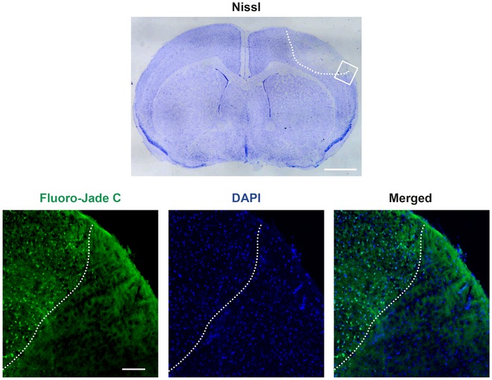
Nissl staining of coronal cryosections (30 μm) corresponding to an animal sacrificed 5 h after ischemic induction reveal a hypochromatic area indicative of neuronal injury in the ipsilateral neocortex (highlighted by a white discontinuous line), compared with equivalent regions of the contralateral hemisphere. Adjacent cryosections were analyzed by Fluoro‐Jade C (green), which specifically stains degenerating neurons. Cell nuclei were stained with DAPI (blue). The interface between the infarcted and non‐infarcted cortical tissue is indicated as before. Scale bar, 1 mm (Nissl); 100 μm (Fluoro‐Jade C).