-
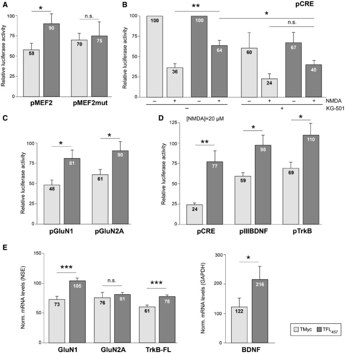
A
Effect of TFL457 on MEF2‐promoter activity. Cultures transfected with pMEF2 (two minimal wild‐type MEF2 elements) or pMEF2mut (mutant) were preincubated with CPPs (25 μM, 30 min) and treated with NMDA (100 μM, 2 h). Means ± SEM of luciferase activities obtained in excitotoxicity, relative to values found in cells treated with same peptide and no NMDA, are presented. Significance was analyzed by Student's t‐test (*P = 0.039; n.s. = non‐significant, P = 0.79; n = 8).
-
B
Effect on CRE‐promoter activity. pCRE contains two minimal CREs. Peptide preincubation was as above with or without KG‐501 (10 μM). Mean ± SEM luciferase activities is given relative to values in cells treated with the same peptide but no NMDA or KG‐501. Differences found in excitotoxicity were analyzed by ANOVA test followed by post hoc Tukey's HSD test (*P = 0.023; **P = 0.003; n.s. = non‐significant, P = 0.20; n = 6).
-
C
Effect on NMDAR‐subunit promoters. Cells transfected with pGluN1 or pGluN2A were treated and analyzed as in panel (A). Significance was analyzed by Student's t‐test (pGluN1, *P = 0.016; pGluN2A, *P = 0.029; n = 14).
-
D
Effects on BDNF promoter III or TrkB promoter. Cells transfected with pIIIBDNF (n = 9), pTrkB (n = 7), or pCRE (n = 5), as a control, were processed as above but using 20 μM NMDA for 4 h. Data are presented and analyzed as in panel (A). Significance was analyzed by Student's t‐test (pCRE, **P = 0.0098; pIIIBDNF, *P = 0.029; pTrkB, *P = 0.033).
-
E
Effects of TFL457 on mRNA levels of CREB/MEF2‐regulated genes. Total RNA was extracted from cultures preincubated with CPPs (25 μM, 30 min) and treated or not with NMDA (100 μM, 4 h). Levels of mRNA were normalized to NSE (genes expressed in neurons, left panel) or GAPDH (neuronal and glial expression, right panel). Means ± SEM of levels obtained in excitotoxicity relative to values found in cells treated with same peptide and no NMDA are presented. Differences found in excitotoxicity were analyzed by ANOVA test followed by post hoc Tukey's HSD test (GluN1, ***P = 0.00096; GluN2A, P = 0.98; TrkB‐FL, ***P = 0.00037; BDNF, *P = 0.045; n = 8).