
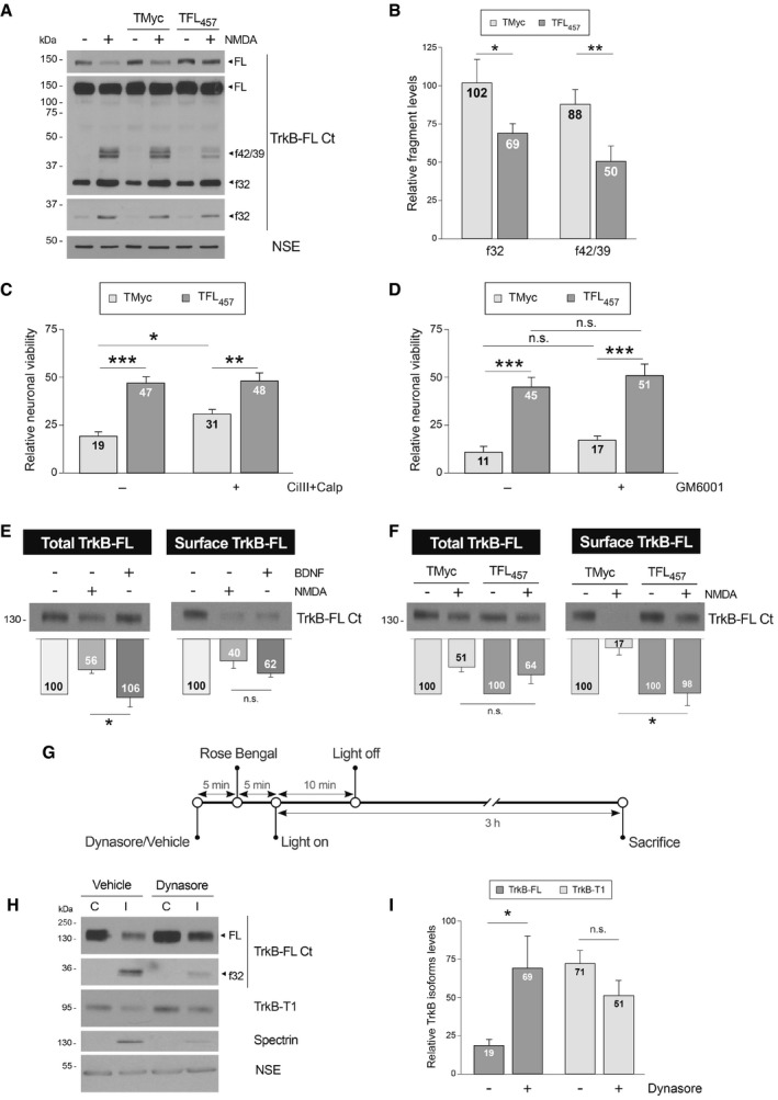
Figure 6. Preservation of TrkB‐FL in the cell surface by TFL457 precedes interference of RIP and calpain processing in excitotoxicity.
-
ATFL457 interference of RIP and calpain processing in excitotoxicity. Cultures were preincubated with peptides (25 μM, 30 min) or left untreated before NMDA addition (2 h). Different exposures are shown to facilitate visualization of FL, f42/39, or f32.
-
BQuantitation of normalized f32 and f42/39 levels. Means ± SEM are shown relative to cultures treated with NMDA and no peptide. Analysis was performed by unpaired Student's t‐test (*P = 0.014, **P = 0.00198; n = 6).
-
CEffect of TFL457 on neuronal viability after calpain inhibition. After 30 min preincubation with calpeptin (Calp, 10 μM) and calpain inhibitor III (CiIII, 10 μM), cultures were treated with CPPs and NMDA (4 h) as before. Means ± SEM relative to those obtained in untreated cultures are given. We performed ANOVA test followed by post hoc Tukey's HSD test (*P = 0.035, **P = 0.0058, ***P = 0.00009; n = 12).
-
DEffect of TFL457 on neuronal viability after metalloproteinase inhibition. Cultures preincubated with GM6001 (10 μM, 30 min) were treated with CPPs and NMDA as before. Analysis was performed as above (without GM6001, ***P = 0.0007; with GM6001, ***P = 0.0006; n = 4).
-
EEffect of NMDA on total and cell‐surface TrkB‐FL levels. Cultures were incubated with NMDA (100 μM) or BDNF (100 ng/ml) for 1 h. Membrane proteins were labeled and purified and compared to corresponding total lysates. TrkB‐FL levels are expressed relative to untreated cells. Mean ± SEM is presented and analyzed by Student's t‐test (*P = 0.027; n.s. = non‐significant, P = 0.1; n = 7).
-
FEffect of TFL457 on the decrease in TrkB‐FL surface levels induced by NMDA. Analysis was performed as before in cells incubated with CPPs (25 μM, 30 min) and NMDA‐treated (1 h). Results obtained in excitotoxic cultures are expressed relative to cells treated with the same peptide but no NMDA. Data are analyzed as in panel (E). Mean ± SEM is presented and analyzed by Student's t‐test (*P = 0.019; n.s. = non‐significant, P = 0.44; n = 4).
-
GExperimental design to study the effect of endocytosis inhibition on TrkB‐FL downregulation in ischemia. Mice were i.p. injected with vehicle or dynasore followed by Rose Bengal. Vessel occlusion and brain damage were induced by cold‐light irradiation. Animals were sacrificed early after damage initiation.
-
HEffect of endocytosis inhibition on TrkB‐FL downregulation. The infarcted area of the ipsilateral hemisphere (I) was compared to the corresponding region of the contralateral area (C). Results from representative mice injected with dynasore or vehicle are shown. Different exposures are presented to facilitate visualization of dynasore effects on TrkB‐FL and f32.
-
IQuantitation of TrkB‐FL and TrkB‐T1 in the infarcted area. Normalized protein levels are expressed relative to those of the corresponding contralateral region. Mean ± SEM is presented and analyzed by Student's t‐test (*P = 0.04; n.s. = non‐significant, P = 0.14; n = 9).
Source data are available online for this figure.