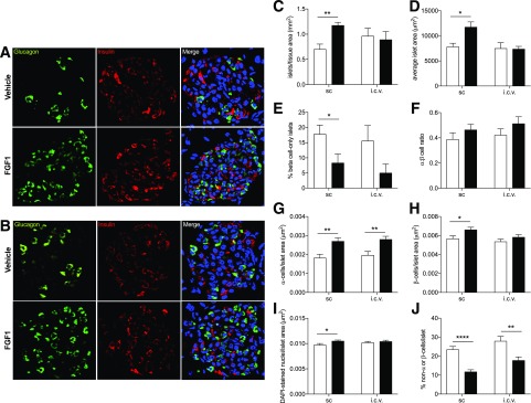
Figure 3.
Chronic central and peripheral FGF1 improved islet microanatomy in db/db mice. Representative images of islet microanatomy after 7 days of FGF1 or vehicle treatment via subcutaneous injections every other day (A) or a single intracerebroventricular (i.c.v.) injection in db/db mice (B). C–J: Histological analysis of representative images from FGF1- (filled bars) and vehicle-treated (open bars) db/db mice. The following parameters were analyzed: islets/tissue area (mm2) (C), average islet area (μm2) (D), percent of β-cell–only islets (E), ratio of α- to β-cells (F), α-cells/islet area (μm2) (G), β-cells/islet area (μm2) (H), DAPI-stained nuclei/islet area (μm2) (I), and percent of non–α-cells or –β-cells/islet (J). Anti-insulin (red), antiglucagon (green), and DAPI (blue) were used for detection of β-cells, α-cells, and nuclei, respectively. Data are expressed as mean ± SEM. P values were determined by unpaired Student t test. *P < 0.05; **P < 0.01; ****P < 0.0001. i.c.v., intracerebroventricular; sc, subcutaneous.

