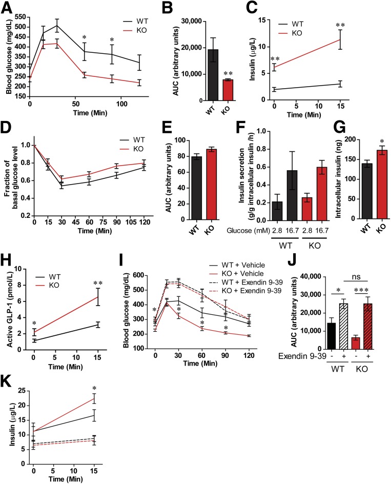
Figure 2.
Loss of Fpr1 in HFD/obese mice improves glucose tolerance and is GLP-1 dependent. A and B: OG-GTT after 10 weeks on an HFD, and area under the curve (AUC) (n = 7–10 mice/group). C: Plasma insulin levels during OG-GTT. D and E: Intraperitoneal insulin tolerance test after 12 weeks on an HFD and AUC (n = 7–10 mice/group). F and G: Glucose-stimulated insulin secretion and intracellular insulin levels in pancreatic islets from HFD WT and Fpr1-KO mice (n = 4 mice/per group, 20 islets/incubation). H: Active GLP-1 levels during the OG-GTT in A (n = 24–25 mice/group). I and J: OG-GTT and corresponding AUC of WT and Fpr1-KO HFD mice after 10 weeks on an HFD and treatment with or without the Glp1r antagonist exendin 9-39 (n = 10–11 mice/group). K: Plasma insulin levels during the OG-GTT in I (n = 10–11 mice/group). Error bars indicate the SEM. ns, not significant. *P < 0.05, **P < 0.001, ***P < 0.0001 by two-way ANOVA and Bonferroni post-test (A and I), one-way ANOVA and Bonferroni post-test (J) or two-tailed t test. Statistical significance is indicated comparing groups at each respective time point. In I and K, significance is indicated for WT vs. Fpr1-KO animals treated with vehicle.

