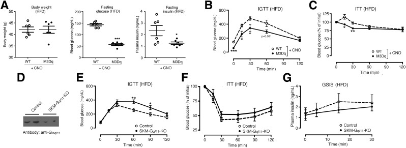
Figure 5.
Metabolic studies with SKM-M3Dq transgenic and SKM-Gq/11 mutant mice maintained on an HFD. A–C: Metabolic studies with SKM-M3Dq mice (M3Dq) and WT control littermates maintained on an HFD for at least 8 weeks. Prior to metabolic testing, CNO (0.25 mg/mL) was added to the drinking water for 1 week. A: Body weight and fasting blood glucose and plasma insulin levels. B: IGTT. Mice were injected with glucose (2 g/kg i.p.). C: ITT. SKM-M3Dq mice (M3Dq) and WT control littermates were injected with insulin (0.75 units/kg i.p.). D–G: Generation and analysis of mice lacking Gαq selectively in SKM (genetic background: Gα11−/−) (SKM-Gq/11 KO mice). D: Representative Western blot indicating that Gαq/11 expression is greatly reduced in SKM (triceps muscle) of SKM-Gq/11 KO mice (genotype: HSA-Cre(ERT2) Gaqflox/floxGa11−/−). Gaqflox/floxGa11−/− mice littermates that lacked the Cre(ERT2) transgene served as control animals. SKM-Gq/11 KO and control littermates consumed an HFD for at least 8 weeks. E: IGTT. Mice were injected with glucose (2 g/kg i.p.). F: ITT. Mice were injected with insulin (0.75 units/kg i.p.). G: Glucose-stimulated insulin secretion (GSIS). Mice were treated with glucose (2 g/kg i.p.), followed by the measurement of plasma insulin levels at the indicated time points. All experiments were carried out with male littermates that were at least 16 weeks old. Data are presented as means ± SEM (6 mice per group). *P < 0.05, **P < 0.01, ***P < 0.001 vs. WT (Student t test).

