Figure 5.
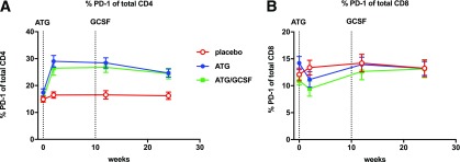
Low-dose ATG enhances PD-1 expression on CD4+ but not CD8+ T cells. Percent of cells expressing PD-1 within the CD4+ (A) or CD8+ (B) T-cell gates are shown, with average measures at the longitudinal time points (0, 2, 12, and 24 weeks) by treatment arm with SDs. Dotted lines denote initiation of ATG and last dose of GCSF. Pairwise comparisons were performed at each time point for placebo vs. ATG (all time points P < 0.001) and vs. ATG/GCSF (all time points P < 0.01).

