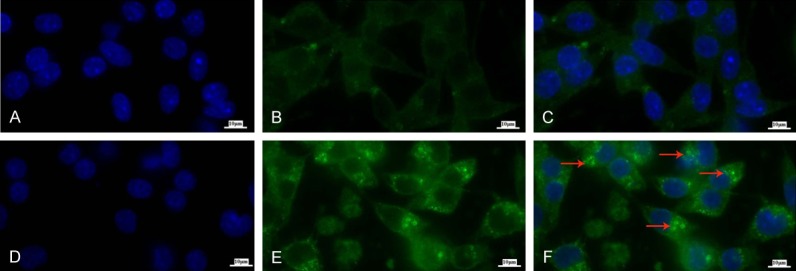
Figure 2.

DSF/Cu increases levels of autophagosome puncta. The cell line MDA-MB-231 was treated in the absence (A-C) or presence (D-F) of DSF/Cu for 24 hours. Cells were harvested, fixed and permeabilized for intracellular LC3B staining. The autophagosomes in the cytoplasm were observed by an immunofluorescence microscope (10×100) (ZEISS AXIOScope. A1). Blue-DAPI for nucleus (A, D), Green-LC3B (B, E) and merged (C, F), autophagosomes as indicated by the red arrows in (F).
