Figure 6.

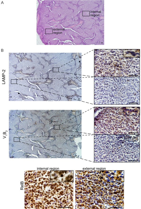
Immunoistaining of Ewing sarcoma xenografts showing the same localization for the indirect markers of acidosis V1B2, LAMP2, and the nuclear localization of RelB, a subunit of the NF-κb transcription complex that can be activated by the TRAF-cIAP destruction complex. (A) H&E staining of a section a representative xenograft of Ewing’s sarcoma model of the control group shown in Figure 1 (scale bar, 200 µm, 4 × objective). We focused our investigation on an external and an internal region of the tumor. (B) Consecutive tissue sections of the section showed in (A), and stained for different antigens. For LAMP2 and V1B2 in the left panel, lower magnification (scale bar 200 µm, 4 × objective), and in the right panels, higher magnification of the specified area unlighted at low magnification by the rectangle (scale bar 20 µm, objective 60 ×). Note in the rectangle showing an enlarged detail, for V-ATPase, the staining is punctuated suggesting a intracellular vesicular localization, for LAMP2 is both punctuated and at the cytoplasmic membrane. For RelB, only high magnification of the two different regions, external and internal are shown (scale bar 20 µm, objective 60 ×). Note in the rectangle showing an enlarged detail that cytoplasm was always positive, whereas nuclei in the internal region were almost stained suggesting the nuclear translocation of the protein, whereas in the external region of the tumor only a small fraction of nuclei were stained.
