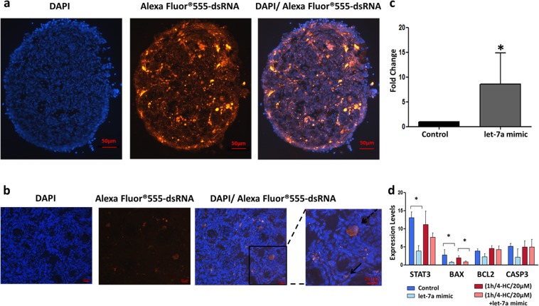
Figure 3.
Evaluation of transfection’s efficiency in PND3 ovaries. (a) Representative images of PND3 ovary, after 2 days of transfection with Alexa Fluor® 555-labeled, dsRNA/ Lipofectamine RNAiMax. PND3 ovarian sections (10 µm) showed nuclear labelling with Hoechst (blue) and Alexa Fluor® 555-labeled, dsRNA (red). The merged images indicate that the dsRNA was been successfully transferred into the ovaries. (b) Magnified images indicate that after transfection the labelled-dsRNA is located mainly in stroma and granulosa cells (bold arrow) and in the oocyte of some follicles (dashed arrow). (c) Expression levels of let-7a after transfection with let-7a mimic using liposome-delivery system. The fold change of let-7a is significantly higher in PND3 ovaries compared to control conditions (N = 3), *p-value < 0.05. The error bars represent the standard error. (d) Expression levels of genes involved in apoptosis in four different groups: control, chemotherapy alone (1 h/4-HC/20 µM), let-7a mimic alone (let-7a mimic), chemotherapy + let-7a mimic ((1 h/4-HC/20 µM) + let-7a mimic). STAT3 and BAX are significantly downregulated in the control + let-7a group (N = 5). BAX is significantly downregulated in the chemo + let7-a group (N = 8).

