Figure 1.
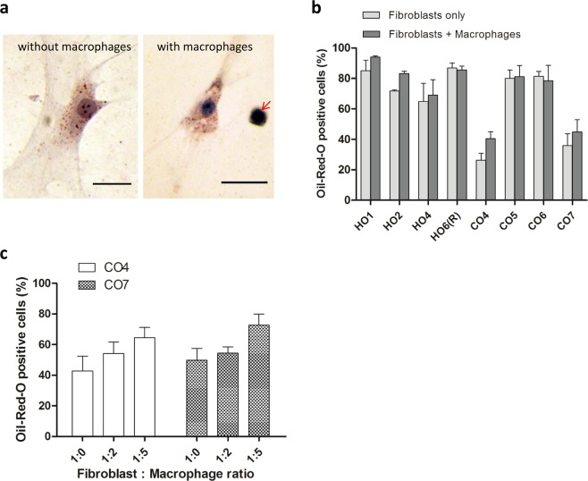
Macrophages do not significantly alter spontaneous lipid vesicle formation in 3D-cultured orbital fibroblasts. (a) Representative Oil-Red-O (ORO) staining of lipid-containing vesicles in orbital fibroblasts in 3D culture for 7 days: HO2 without macrophages (left) and CO6 with macrophages (right, arrow), at a 1:1 fibroblast:macrophage ratio. Scale bar, 25 μm. (b) Macrophages did not alter spontaneous lipid vesicle formation in 3D culture. GO (HOs) or control (COs) fibroblasts (0.74 × 10⁵ cells/ml) were cultured with macrophages at a 1:1 fibroblast:macrophage ratio in the presence of 10% serum for 7 days. The fraction of ORO positive cells is shown as mean ± SEM (n = 3, no statistical difference between with and without macrophages). (c) ORO positive cells in control fibroblast lines, CO4 and CO7, did not significantly increase as the number of macrophages increased (mean ± SEM; n = 4, p = 0.118 (CO4) and p = 0.123 (CO7), Kruskal-Wallis test).

