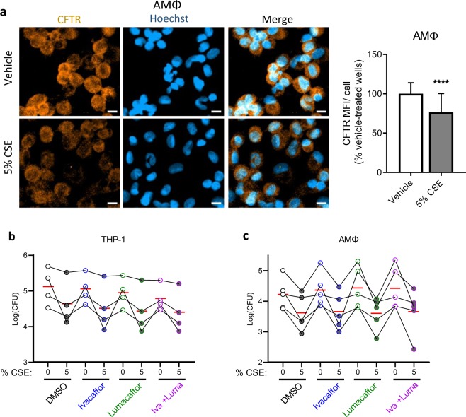
Figure 2.
CSE decreases CFTR expression while CFTR modulators fail to rescue phagocytosis. (a) Primary AMΦ from healthy donors were plated overnight onto glass slide chambers. The following morning they were treated with vehicle or 5% CSE for one hour. They were then washed, fixed in methanol, and stained for CFTR and counterstained with Hoechst. Three 20x fields in three separate wells for each condition were imaged, mean orange fluorescence was calculated and divided by the number of cell nuclei/field then normalized to vehicle-treated wells. Mean and standard deviations from five independent experiments from different donors with 6–9 fields imaged from each are graphed, ****p < 0.0001 by Mann-Whitney test. Scale bars = 10 μm. (b) Phagocytosis assay was performed with THP-1 cells as in Fig. 1 except cells were pre-treated with DMSO, ivacaftor 30 nM, lumacaftor 3 μM or the combination of the two for 48 hrs prior to assay, after differentiation with PMA. Points with connecting lines represent means from four individual experiments, each performed in triplicate. Red horizontal lines indicate mean of the four experiments. Linear models revealed that all three treatments reduced log10 CFU (ivacaftor: p = 0.08; lumacaftor: p < 0.01; 5% CSE: p < 0.001) in THP-1 cells. (c) In AMΦ, only 5% CSE was associated with significant reduction in phagocytosis (p < 0.001).

