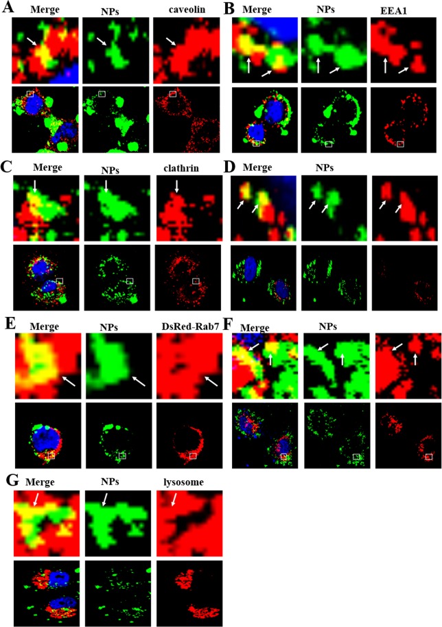
Figure 2.
Confocal images of endocytosis pathways. In MCF-7 cells, the NPs enter the cells through clathrin-dependent and caveolin-dependent endocytosis (A–C) after treatment with 1 mg/mL coumarin-6-labeled NPs for 3 h. Clathrin, caveolin and EEA1 were detected by primary antibodies against clathrin, caveolin and EEA1. (D–F) DsRed-Rab5, 7 and 9 cells were incubated with 1 mg/mL coumarin-6-labeled NPs for 3 h. (F) 1 mg/mL coumarin-6-labeled NPs transfected MCF-7 cells for 3 h and detected by Lyso-Tracker Red probes for 30 min. The above images are the enlarged ones in the white collar on the underside images. Scale bars: 10 μm.

