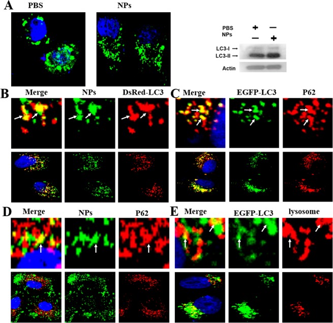
Figure 4.
Confocal microscopy studies autophagy induced by NPs. (A) EGFP-LC3 cells were cultured with 1 mg/mL Coumarin-6-labeled NPs for 3 h. Western blotting of the cells cultured in (A). (B) DsRed-LC3 cells were cultured with 1 mg/mL Coumarin-6-labeled NPs for 3 h. (C) MCF-7 cells were cultured with 1 mg/mL NPs for 3 h, and P62 was detected with a primary antibody against P62. (D) The autophagosomes fused with lysosomes (arrows). (E) EGFP-LC3 cells were cultured with 1 mg/mL NPs for 3 h and Lyso-Tracker Red probes for 30 min. The above images are the enlarged ones in the white collar on the underside images. Scale bars: 10 μm.

