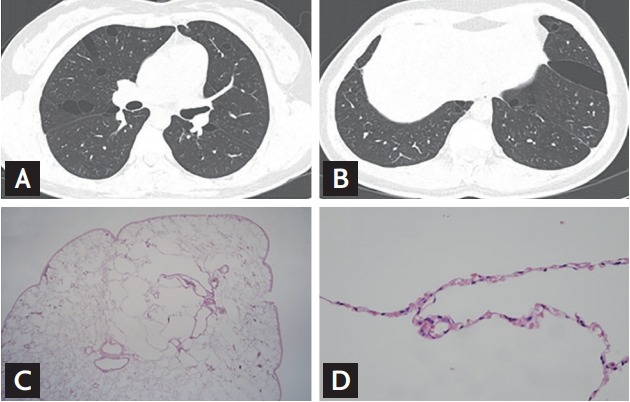
Figure 2.

Examples of chest computed tomography and histologic findings of the study patients. The pulmonary cysts in Birt-Hogg-Dubé (BHD) are distributed in predominantly lower, peripheral and subpleural regions of the lungs (A: upper lobes; B: lower lobes). Lung specimens present multiple small intraparenchymal cysts rimmed by thin fibrous walls or normal pulmonary parenchyma. (C: ×40; D: ×400).
