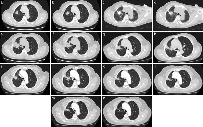
Figure 1.

(a, b) A 48‐year‐old female was diagnosed with a right upper lobe lung mass and left renal mass. She was confirmed to have a primary lung adenocarcinoma with renal metastatic carcinoma on biopsy. CT showedithe primary lung tumor was 2.5 cm in diameter. (c, d) Puncture of the ablative antenna into the tumor. (e, f) One month after microwave ablation (MWA). (g, h) Four months after MWA. (i, j) Seven months after MWA. (k, l) Ten months after MWA. (m, n) Thirteen months after MWA. The tumor hasshrunk and fibrosis has developed, An irregular cavity has also formed.
