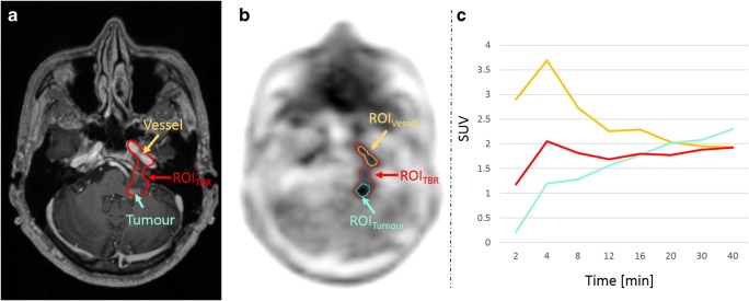
Fig. 3.
a T1-weighted MRI with a contrast-enhancing lesion (cyan arrow) and the arteria carotis interna (orange arrow). b ROITBR delineation of a tumour in the PET image after AC using the model-based approach. In this case, the ROITBR additionally includes parts of the arteria carotis interna. c TACs extracted from ROITBR (red) and from two manually drawn ROIs in the tumour (ROITumour, cyan) and the arteria carotis interna (ROIVessel, orange). The TAC extracted from ROITBR is a mixture of the TAC extracted from ROITumour and ROIVessel

