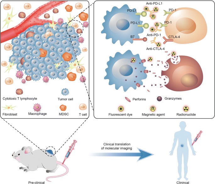
Fig. 1.
Targeted molecular imaging of immune checkpoints from preclinical to clinical studies. In tumor micro-envirenment, radionuclide, fluorescent dye, or magnetic agent labeled monoclonal antibodies as anti-PD-L1, anti-PD-1, anti-CTLA4 et al were performed using SPECT, PET/CT, MRI, or optical imaging. Cytotoxic T lymphocytes were activated by immune checkpoint blocking treatment causing a higher releasing of granzyme B; radionuclide-labeled granzyme B was utilized as a target for PET imaging

