Fig. 2.
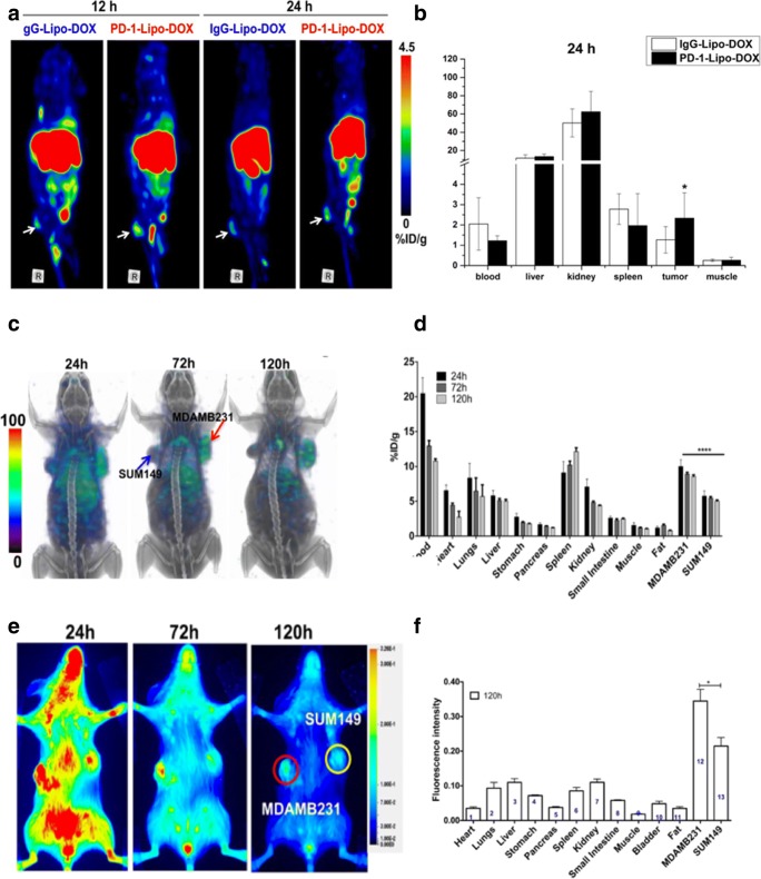
Multimodality molecular imaging of PD-1/PD-L1 expressing cancer. a, b PET images of 4T1 mammary tumor-bearing mice at 12 and 24 h postinjection of PD-1-Liposome-DOX-DOTA-64Cu, and biodistribution of PD-1-Liposome-DOX-DOTA-64Cu in 4 T1 tumors 24 h postinjection. c, d SPECT images were acquired at 24, 72, and 120 h after the injection with 14.8 MBq (400 uCi) of 111In-PD-L1-mAb. The SPECT images showed the higher intensity biodistribution of 111In-PD-L1-mAb in the MDA-MB-231 tumor compared to SUM149 tumor in the same tumor-bearing mice, and ex vivo biodistribution analysis of [111In] radioactive tracer intensity in the different tissues at 24 h, 72 h, and 120 h postinjection. e, f Optical images showed specific fluorescence biodistribution of NIR-PD-L1-mAb in the MDA-MB-231 tumor compared to SUM149 tumor in the same tumor-bearing mice, and the ex vivo biodistribution analysis of fluorescence intensity in the different tissues at 120 h postinjection (a and b from Du, with permission of [14]. c–f [15] by Chatterjee S is licensed under CC BY 3.0)

