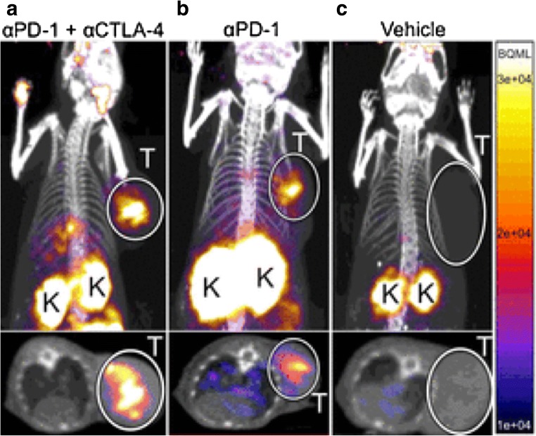
Fig. 4.
PET imaging of granzyme B following immune checkpoint blocking. The coronal and axial maximal intensity projection imaging of PET images of anti-PD-1 and anti-CTLA-4 combination-treated (a), anti-PD-1-treated (b), and vehicle-treated (c) colon tumor-bearing mice acquired 1 h postinjection of 68Ga-NOTA-GZP. The PET imaging showed high radionuclide signal intensity with 68Ga-NOTA-GZP in tumor with combination treatment of anti-CTLA-4 and anti-PD-1 antibodies. T tumors, K kidneys. (From Larimer BM, with permission of [35])

