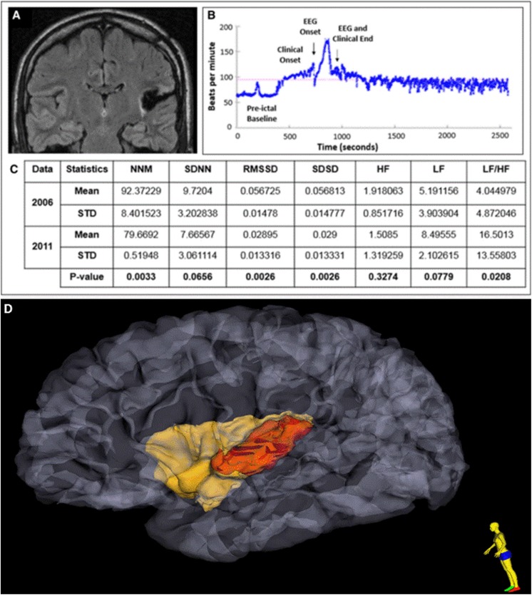
Figure 8.
A, Postoperative MRI FLAIR sequence shows evidence of a left posterior temporo-insular resection cavity with surrounding gliosis in a patient with later SUDEP. B, Heart rate plots show ictal sinus tachycardia, followed by sustained postictal sinus tachycardia lasting at least 25 minutes after a nonfatal generalized convulsive seizure. C, Heart rate time and frequency domain parameters calculated during the presurgery (2006) and postsurgery (2011) epilepsy monitoring unit (EMU) evaluations and the results from generalized estimating equation (GEE) analysis. D Extent of insular resection and damage, after 3-dimensional reconstruction of pre- and postoperative MRI is delineated in red. MRI indicates magnetic resonance imaging; FLAIR, fluid-attenuated inversion recovery; SUDEP, sudden unexpected death in epilepsy; MNN, mean of normal to normal heart beats; SDNN, standard deviation of normal to normal heart beats.

