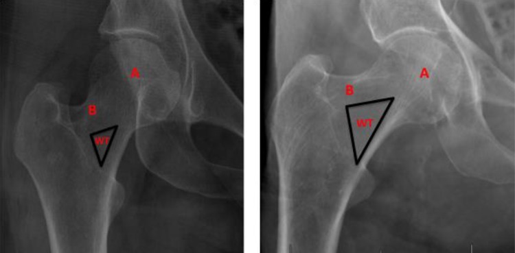
Figure 1.
(Left) Right hip x-ray of a 30-year-old female. (Right) Right hip x-ray of a 98-year-old male. Note that the size of Ward triangle (WT) is significantly larger in the right image compared to the left image and that there is greater degeneration of principle compressive trabeculae (A) and principle tensile trabeculae (B) in the right image compared to the left image.

