. 2019 May 22;10(26):6503–6507. doi: 10.1039/c9sc00531e

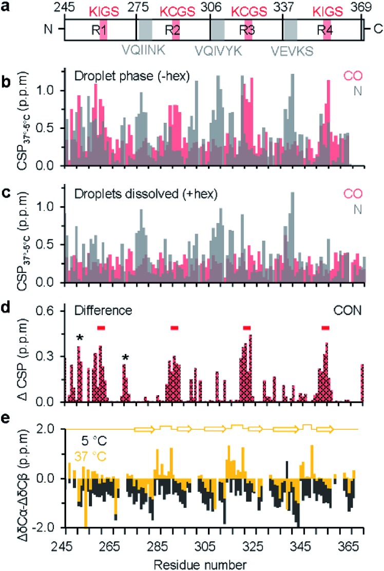
Fig. 2. Residue-specific analysis of LLPS-induced changes in chemical environment and local structure in the repeat-region of tau. (a) Domain representation of K18. Pseudo-repeats are shown as R1–R4, hexapeptides are marked in grey, KXGS motifs in red. (b) Chemical shift perturbation (CSP) of CO (red) and N (grey) from the dispersed monomeric (5 °C) to the droplet state (37 °C). (c) CSP of CO and N from the dispersed monomeric state (5 °C) to the dispersed monomeric state at 37 °C (i.e. in the presence of 1,6-hexanediol), revealing temperature-induced changes in N chemical shifts. (d) Averaged CSP (ΔCSP) of CO and N in K18 between the droplets state (without 1,6-hexanediol at 37 °C) and after droplet dissolution (with 1,6-hexanediol at 37 °C). The four KXGS motifs (red bars), as well as the proline-rich region and the PGGG motif (asterisk) in R1 display pronounced ΔCSP. (e) Secondary structure propensity of K18 in the monomeric dispersed state at 5 °C (black) and the LLPS-phase at 37 °C (yellow). At 37 °C, secondary chemical shifts suggest the presence of β-hairpin-like conformations in repeats R2, R3 and R4 (illustrated by a cartoon diagram on top).

Fig. 2