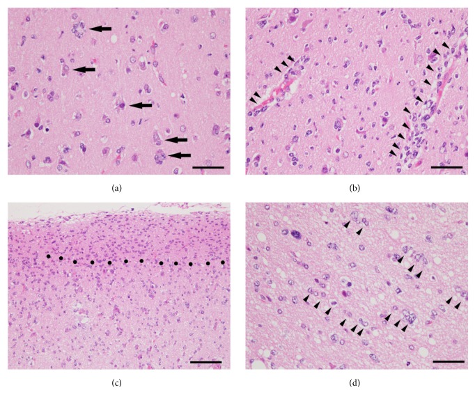
Figure 2.
Specific histomorphological patterns of diffuse invasion, so-called “secondary structures of Scherer” in glioblastoma. As a rule, glioma cells migrate along existing brain structures such as brain parenchyma, blood vessels, white matter tracts, and subpial spaces. The secondary structures of Scherer are referred to four criteria as (a) perineuronal satellitosis (indicated by arrows), (b) perivascular satellitosis (indicated by arrow heads), (c) subpial spread (region above black dots), and (d) invasion along the white matter tracts (indicated by arrow heads). Hematoxylin and eosin staining. Scale bars in (a), (b), and (d) are 50 μm; scale bar in (c), 100 μm.

