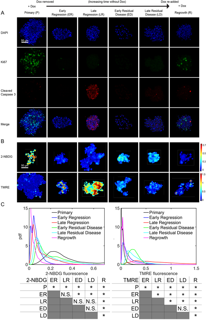
Figure 4: Tumor metabolism reprograms during the different stages of a tumor’s life cycle.
Mammospheres stained for either Ki67 and cleaved caspase 3 (CC3), 2-NBDG, or TMRE were imaged from cultures with Dox in the media (Primary, P), without Dox in the media for 2 days prior to imaging (Early Regression, ER), without Dox in the media for 4 days prior to imaging (Late Regression, LR), without Dox in the media for 14 days prior to imaging (Early Residual Disease, ED), and without Dox in the media for 28 days prior to imaging (Late Residual Disease, LD). Cells were also cultured without Dox for 21 days followed by Dox added back in for 7 days (Regrowth, R). (A) Representative image planes of DAPI, Ki67, cleaved caspase 3 (CC3) staining. (B) Representative mean projection images of 2-NBDG and TMRE intensities. (C) Probability density functions of 2-NBDG and TMRE intensities from the 3D mammospheres show significantly decreased 2-NBDG and significantly increased TMRE uptake after Dox withdrawal. After re-addition of Dox to dormant tumors, 2-NBDG uptake remains significantly decreased. Significant comparisons between time points for both 2-NBDG and TMRE are included. For all groups n=10 mammospheres. (* is p<0.05, N.S. is not significant).

