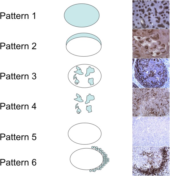
Fig. 1.

Patterns of CD21 and CD23 expression by the follicular dendritic cells. Pattern 1 describes diffuse staining of FDCs in the germinal center while pattern 2 demonstrates the peripheral staining in a “crescetic” fashion. Pattern 3 shows broken networks of the FDCs while the follicular outline is preserved. In pattern 4, the follicular outline is lost. Pattern 5 shows lack of stain and, in pattern 6, there is staining of FDC extensions around the individual mantle zone B cells
