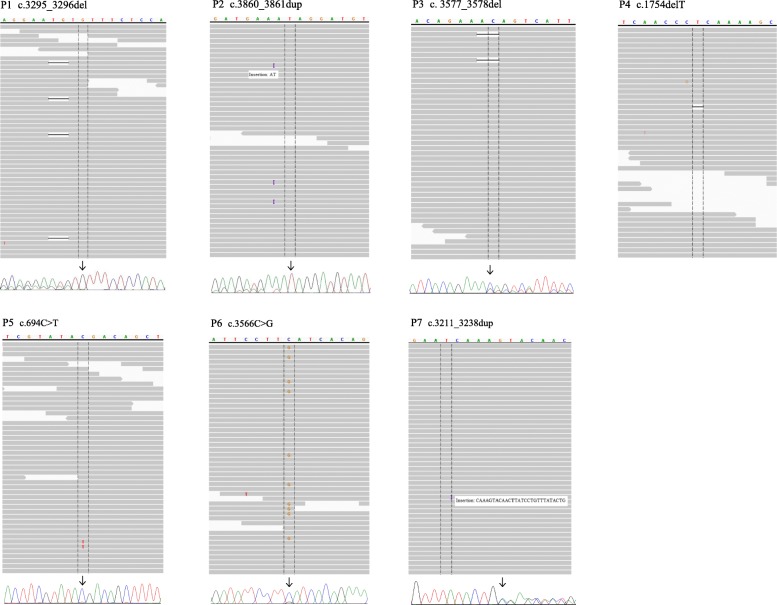
Fig. 1.
Visual verification of variants with Integrative Genomic Viewer (IGV) and sequencing chromatogram with secondary confirmation test results. Variants with low fractions in IGV reflect NGS results from analyzing peripheral blood. The corresponding sequencing chromatograms are the results of MEMO-PCR of peripheral blood for P1 and P2, conventional PCR of polyp tissue for P3 and P5, and conventional PCR of peripheral blood for P6 and P7

