
Fig. 4. The cleaved NLRP1B N terminus is sufficient to induce protein degradation.
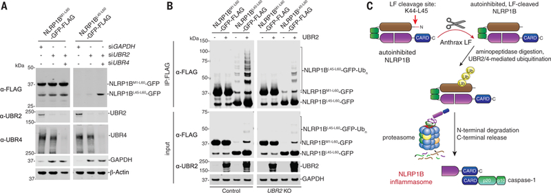
(A) HEK 293T cells were transfected with the indicated small interfering RNAs, incubated for 24 hours, then transfected with NLRP1BM1−60- or NLRP1BL45−60-GFP-FLAG fusion constructs (0.05 μg) for an additional 24 hours. Lysates were then evaluated by immuno-blotting. (B) WT or UBR2 KO HEK 293T cells were transfected with the indicated NLRP1BM1−60- or NLRP1BL45−60-GFP-FLAG fusion constructs (0.5 μg) and UBR2 (1.5 μg) and incubated for 24 hours. Lysates were harvested, immunoprecipitated with anti-FLAG agarose beads, and evaluated by immunoblotting. Data are representative of three or more independent experiments. (C) Proposed model of LT-induced NLRP1B inflammasome activation.