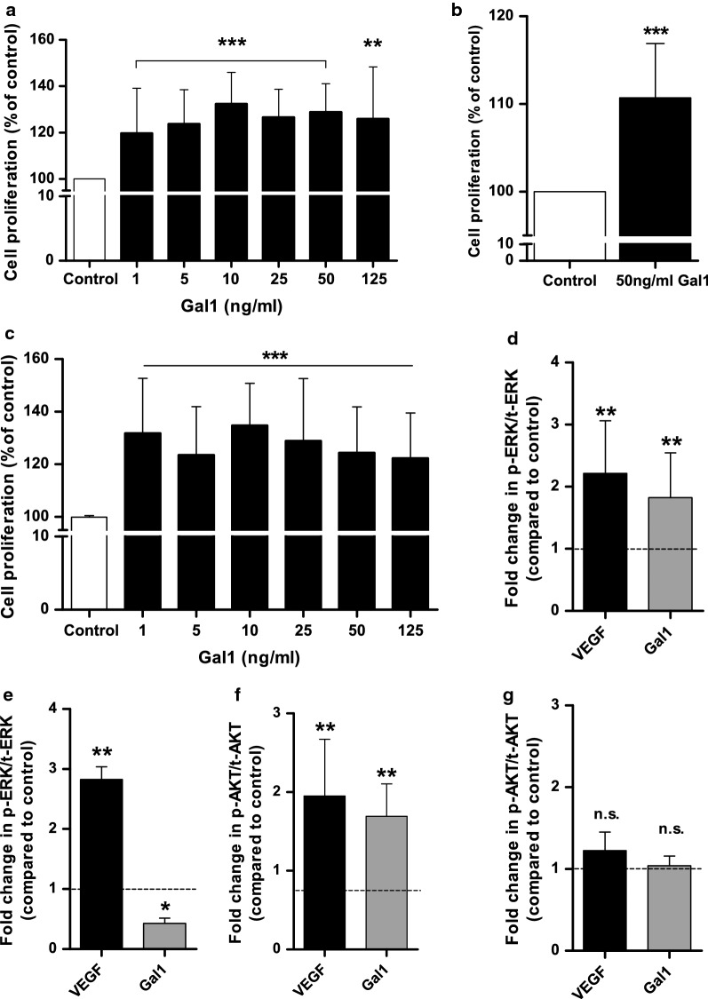
Fig. 2.
Gal1 induces HOMEC proliferation and activation of ERK1/2 and AKT. Cells were seeded in 2% gelatin pre-coated 96 well plates at a density of 10,000 cells/well in starvation media containing 2% FCS. a–c After overnight incubation, cells were treated ± Gal1 at various concentrations and incubated for 48 h. a WST-1 assay was used to assess cellular proliferation based on absorbance using a PHERAstar BMG plate-reader at 450 nm (n = 20). b Cell proliferation was examined at 50 ng/ml of Gal1 (CyQUANT). A commercially available CyQUANT reagent was used to assess cell proliferation after 48 h treatment based on fluorescence intensity using a FLUOstar BMG plate-reader at Ex/Em: 485/530 nm (n = 20). c A commercially available BrdU reagent was added to the wells for the last 24 h incubation and cellular proliferation was assessed (according to the manufacturer’s instructions) at 48 h based on absorbance using a SpectraMax plate-reader at 450/550 nm (n = 15). Results are mean ± SD and shown as percentage of the control, **p < 0.01 and ***p < 0.001 vs control (100%). After overnight incubation, cells were treated ± 50 ng/ml of Gal1 or 20 ng/ml of VEGF and incubated for 4 or 10 min. ERK1/2 (d, e) and AKT (f, g) phosphorylation was examined after 4 min (d, f) and 10 min (e, g) treatments. Commercially available cell-based ELISAs were used for the determination of ERK1/2 and AKT (S473) phosphorylation level. The ELISA experiments were carried out in quadruplets on two cell batches. The data show fold change in phosho-ERK1/2/AKT relative to total ERK1/2/AKT (compared to control). Results are mean ± SD, n.s., *p < 0.05, **p < 0.01 vs control (dotted lines); n = 4–6. n.s. denotes not significant vs control

