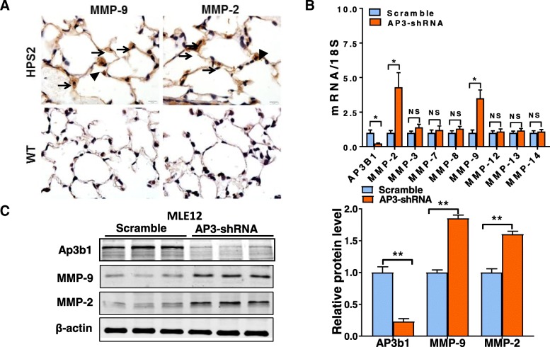
Fig. 2.
HPS2 deficiency leads to increased MMP expression in lung epithelial cells. a Immunohistochemical staining for MMP-2 and MMP-9 (brown staining) in the lungs of wild-type and HPS2 mice. Staining is not detected in the lungs of wild-type mice and alveolar epithelial type II cells are difficult to visualize due to normal size lamellar bodies. Expression of MMP-2 and MMP-9 appears to be increased most significantly in alveolar epithelial type II cells, as indicated by high intensity brown staining in cells containing prominent lamellar bodies (arrows). Brown staining was also evident in some alveolar macrophages (arrow heads) and in the interstitial space. b, c shRNA knockdown of the Ap3b1 gene in MLE12 cells leads to a marked upregulation in transcript (n = 4, per group) and protein levels for MMP-2 and MMP-9. Immunoblot is representative of at least two different blots and densitometry analyses (bar graphs) (n = 5 per group, *p < 0.05, HPS2 vs. control). Data are expressed as mean ± SE, and statistical significance was assessed using a Student’s unpaired t test

