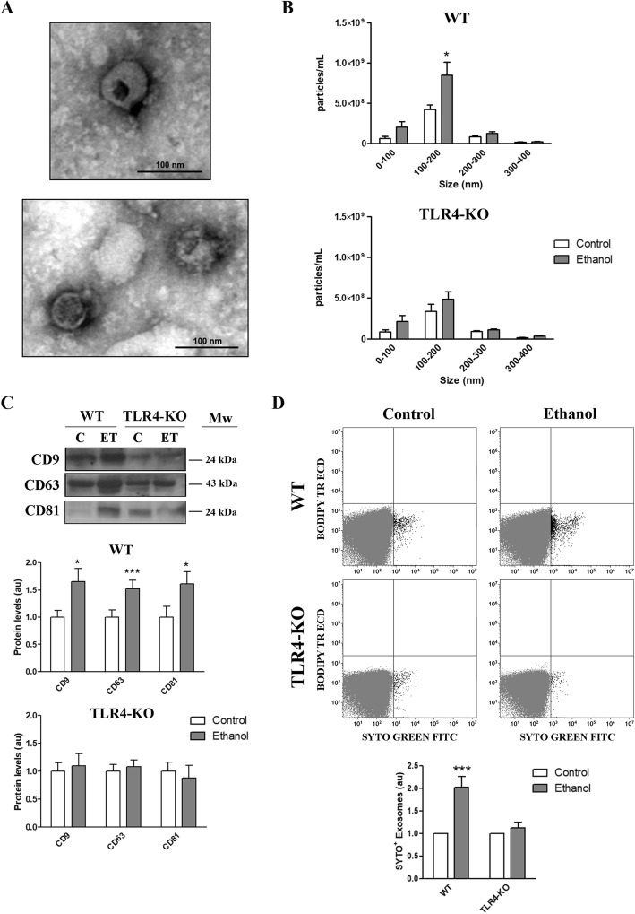
Fig. 1.
Characterization of the EVs secreted by the untreated and ethanol-treated WT and TLR4-KO astrocytes. a Electron microscopy images of astrocyte-derived EVs. Scale bar, 100 nm. b Measurement of the absolute size range and concentration of the microvesicles derived from astrocytes by the nanoparticles tracking analysis. c Immunoblot analysis and quantification of CD9, CD63, and CD81 in astrocyte-derived EVs. A representative immunoblot for each protein and their molecular weight are shown. 40 μg/μL of protein were loaded in each lane. d Flow cytometry analysis and quantification of the astrocyte-derived EVs stained with green-RNA-binding fluorophore SYTO (SYTO+ events are black-colored). The WT and TLR4-KO astroglial cells were treated with ethanol (40 mM) for 24 h. Data represent mean ± SEM, n = 5–7 independent experiments. *p < 0.05 and ***p < 0.001 compared to their respective control group, according to the two-way ANOVA followed by Bonferroni’s post-hoc test

