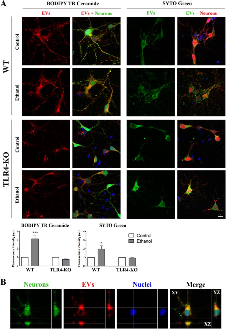
Fig. 5.

Analysis of the internalization of the astrocyte-derived EVs in naïve cortical neurons. Neurons were incubated overnight with 9–10 μg/μL of the fresh astrocyte-derived EVs. a Fluorescence intensity of the red-fluorophore lipid-binding Bodipy or the green-RNA-binding fluorophore SYTO astrocyte-derived EVs internalized by green-stained (Cell Tracker) or red (ACTB-Ds Red) naïve cortical neurons, respectively. A representative photomicrograph of each condition is shown. Scale bar, 10 μm. EVs were obtained from the WT and TLR4-KO astroglial cells treated with ethanol (40 mM) for 24 h. Bar graphs represent the mean ± SEM of the data from three different fields per condition from six distinct cultures. b Confocal microscopy images showing that EVs are internalized in the neuronal cytoplasm, as demonstrated using the xyz axes projections. Green, red, and blue fluorescence represent the cell tracker, the EVs stained with Bodipy, and nuclei staining, respectively. *p < 0.05 and ***p < 0.001 compared to their respective control group, according to the two-way ANOVA followed by Bonferroni’s post-hoc test
