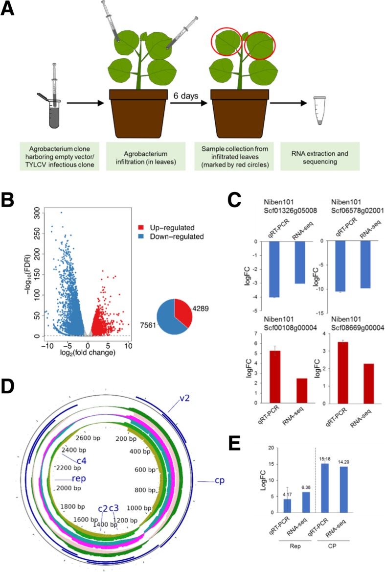
Fig. 1.

Transcriptional changes upon local infection by TYLCV in N. benthamiana. a Schematic representation of the experimental design. b Differentially expressed genes in the local infection are shown by volcano plot. The x-axis shows the log2 transformed gene expression fold change between infected and control samples. The y-axis indicates the negative log10 transformed adjusted p-values (FDR) of the differential expression test calculated by R package edgeR. The up-regulated and down-regulated genes are represented by red and blue dots, respectively. Pie chart shows the number of up−/down-regulated genes. c Validation of selected DEGs by qPCR. Values are the average of three biological replicates, relative to mock. NbACT was used as the normalizer. d Mapping of viral reads to the TYLCV genome. Three biological replicates (inner concentric circles) are represented; the upper side of each circle represents the virion (+) strand; the lower side of each circle represents the complementary (−) strand. ORFs are depicted in blue. Please note that the accumulated reads in the area containing the CP and V2 ORFs have been trimmed; the numbers of total reads are shown in Additional file 7: Table S2. e Validation of the expression of Rep and CP by qPCR. Expression values are relative to NbACT
