Fig. 1.

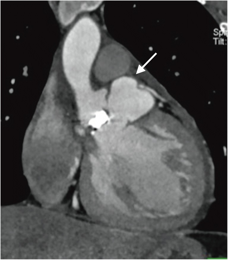
Pseudoaneurysms of the left ventricular outflow tract in patient after aortic valve replacement (arrow). ECG-gated computed tomography, multiplanar reconstruction, frontal oblique view

Pseudoaneurysms of the left ventricular outflow tract in patient after aortic valve replacement (arrow). ECG-gated computed tomography, multiplanar reconstruction, frontal oblique view